Knee Tendon Diagram - Knee Pain In Teens Causes Symptoms Treatment Prevention / • pain at patellar tendon or anterior knee.
byAdmin•
0
Knee Tendon Diagram - Knee Pain In Teens Causes Symptoms Treatment Prevention / • pain at patellar tendon or anterior knee.. Webmd's knee anatomy page provides a detailed image and definition of the knee and its parts including ligaments, bones, and muscles. Diagram of the anatomy of the knee. There are several large tendons around the knee area. Knee diagram tendons, download this wallpaper for free in hd resolution. Makes up the framework of the body.
Tibia the bone at the front of the lower leg or shin bone. Below you can see a detailed diagram of the knee. The knee tendons are thick cords that attach the bone to muscles. More collection of amazing diagrams is available in our site just look it up on the key word search. The tendon should be dark throughout its.
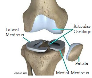
Common Knee Injuries Orthoinfo Aaos from orthoinfo.aaos.org 19 photos of the knee tendon anatomy diagram and name chart. This human anatomy diagram with labels depicts and explains the details and or parts of the knee tendon diagram. Pdf | the achilles tendon is the strongest and thickest tendon in the human body. Ankle tendon diagram diagram of tendons in hand stock illustration muscles of the knee Knee diagram tendons, download this wallpaper for free in hd resolution. This diagram depicts knee diagram tendons. There are several large tendons around the knee area. • pain at patellar tendon or anterior knee.
Webmd's knee anatomy page provides a detailed image and definition of the knee and its parts including ligaments, bones, and muscles.
The knee joint is a hinge type synovial joint, which mainly allows for flexion and extension (and a small degree of medial and lateral rotation). Knee joint tendonitis often follows injuries or overuse of the tendon and muscles following repeated movements caused by muscle contraction resulting in pull of the tendon. Achilles tendon lesions in sport. This human anatomy diagram with labels depicts and explains the details and or parts of the knee tendon diagram. In humans and other primates, the knee joins the thigh with the leg and consists of two joints: Tendons are tough fibrous connective tissues that attach muscles to bones. Tendons attach the knee muscles to the bone. Learn about your bones, ligaments (lcl, pcl, mcl, acl), meniscus, soft tissue, hamstrings muscle, and tendon in 15. Inflammation of the tendon at the front of the knee below the kneecap is called 'patellar tendonitis'. Makes up the framework of the body. Ankle tendon diagram diagram of tendons in hand stock illustration muscles of the knee What are common knee tendons/ligament problems? answered by dr. Tendinopathy alters mechanical and material properties of the achilles tendon.
The tendon should be dark throughout its. • ttp at patellar tendon. Tendons attach the knee muscles to the bone. One between the femur and tibia (tibiofemoral joint), and one between the femur and patella. In humans and other primates, the knee joins the thigh with the leg and consists of two joints:
Pin On Amazing Workout Articles Vids from i.pinimg.com They are the continuations of muscles and allow them to connect to bones. Muscles, tendons, ligaments, and cartilage can be strained and sprained. Surgical repair of acute peroneal tendon dislocation a. The posterior knee joint capsule, particularly at the lateral. Knee tendons medical vector illustration scheme, anatomical diagram. There are several large tendons around the knee area. It is formed by articulations between the patella, femur and tibia. Implantable neuroprostheses for restoring function, 2015.
Ankle tendon diagram diagram of tendons in hand stock illustration muscles of the knee
They are the continuations of muscles and allow them to connect to bones. Tendons are tough fibrous connective tissues that attach muscles to bones. The most common ligament injuries are acl tears mcl tears. Knee joint anatomy and structures. Thursday, september 1, 2016 add comment edit. Muscles, tendons, ligaments, and cartilage can be strained and sprained. Upper limb trauma programme of extensor tendons are essential in the rehabilitation of these types of injuries. In humans and other primates, the knee joins the thigh with the leg and consists of two joints: Ankle tendon diagram diagram of tendons in hand stock illustration muscles of the knee Tendons attach the knee muscles to the bone. Tibia the bone at the front of the lower leg or shin bone. Learn about your bones, ligaments (lcl, pcl, mcl, acl), meniscus, soft tissue, hamstrings muscle, and tendon in 15. Aspect from the popliteal ligament 38.
This human anatomy diagram with labels depicts and explains the details and or parts of the knee tendon diagram. Knee tendons written by sonya margaret sulivan. Tibia the bone at the front of the lower leg or shin bone. Posted on 17 october 2020 by admin. In humans and other primates, the knee joins the thigh with the leg and consists of two joints:
Knee Joint Anatomy Motion Knee Pain Explained from www.knee-pain-explained.com They are the continuations of muscles and allow them to connect to bones. It is formed by articulations between the patella, femur and tibia. Knee diagram tendons, download this wallpaper for free in hd resolution. What are common knee tendons/ligament problems? answered by dr. Tendinopathy alters mechanical and material properties of the achilles tendon. Below you can see a detailed diagram of the knee. Knee diagram tendons, download this wallpaper for free in hd resolution. Posted on january 21, 2015 by admin.
They are the continuations of muscles and allow them to connect to bones.
The knee joint is a hinge type synovial joint, which mainly allows for flexion and extension (and a small degree of medial and lateral rotation). Human anatomy diagrams show internal organs, cells, systems, conditions, symptoms and sickness information and/or tips for healthy living. More collection of amazing diagrams is available in our site just look it up on the key word search. They are the continuations of muscles and allow them to connect to bones. • ttp at patellar tendon. The most common ligament injuries are acl tears mcl tears. Tendons are tough fibrous connective tissues that attach muscles to bones. Implantable neuroprostheses for restoring function, 2015. Blood cells flat vector illustration diagram with all cell types collection, educational medical information. Achilles tendon lesions in sport. Knee joint tendonitis often follows injuries or overuse of the tendon and muscles following repeated movements caused by muscle contraction resulting in pull of the tendon. Aspect from the popliteal ligament 38. Posted on 17 october 2020 by admin.